“The breast tissue is heterogenously dense, which may obscure small masses.”
You may have read this in your recent mammogram report. The word “obscure” does not exactly help bring peace of mind when screening for breast cancer, the most common cancer for women in the US and the second-leading cause of cancer death after lung cancer. The good news is that breast cancer screening is an area where technology is evolving quickly, and one of the most promising newer tools is contrast-enhanced mammography (CEM).
If you’ve recently been told you have dense breasts, or if you’ve ever had an unclear mammogram, there are better ways we can detect breast cancer for you.
What is a contrast-enhanced mammogram?
A contrast-enhanced mammogram is a type of breast imaging that combines:
- A standard mammogram
- A small amount of intravenous (IV) contrast dye
The contrast dye helps highlight areas of increased blood flow. Since cancers often develop new blood vessels, this technique can make suspicious areas more visible than on standard imaging alone.
Think of it as adding a “highlighting effect” to a traditional mammogram.
Why is this important for women with dense breasts?
Dense breast tissue is very common and completely normal—but it can make cancer harder to detect on routine mammograms. On imaging, both dense tissue and tumors can appear white, making it difficult to distinguish between the two.
This is why many patients with dense breasts are offered additional screening, such as ultrasound or MRI. However, newer evidence suggests that CEM may outperform ultrasound in detecting cancers in this group.
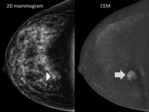
Photo above: Left shows dense breast tissue on a routine 2D mammogram. Right shows image quality improvement with CEM
What does the latest research show?
The BRAID Trial (2025)
The most important study to date is the BRAID randomized controlled trial, which directly compared contrast-enhanced mammography with automated breast ultrasound in women with dense breasts and normal mammograms.
Here’s what researchers found:
- CEM detected 19.2 cancers per 1,000 women, compared to 4.2 per 1,000 with ultrasound
- This represents nearly a fourfold increase in cancer detection
- CEM detected more invasive cancers (15.7 vs. 4.2 per 1,000)
- The cancers found with CEM were about half the size of those found with ultrasound
- Ultrasound did not detect any cases of DCIS (a very early form of breast cancer), while CEM detected several
These findings suggest that CEM may not only detect more cancers—but detect them earlier, when they are more treatable.
There were some trade-offs:
- Higher recall rates with CEM (9.7% vs. 4.0%)
- Small risk of contrast reactions (mostly mild, rarely severe)
Additional Supporting Evidence
A 2025 study published in Radiology compared three approaches:
- Mammography alone
- Mammography + ultrasound
- Contrast-enhanced mammography
Results showed:
- CEM detected 17.1 cancers per 1,000 women
- Mammography + ultrasound detected 8.5 per 1,000
- Mammography alone detected 2.1 per 1,000
Importantly, CEM also had a higher positive predictive value (45% vs. 28%), meaning fewer unnecessary biopsies were performed per cancer found.
How accurate is CEM overall?
A large meta-analysis including more than 10,000 patients found:
- Sensitivity: ~95% (very strong ability to detect cancer)
- Specificity: ~78% (moderate rate of false positives)
In women with dense breasts, these numbers remained similarly strong.
For comparison, adding ultrasound to mammography typically identifies only 2–5 additional cancers per 1,000 women—significantly fewer than what is seen with CEM.
What are the benefits of CEM?
Based on current evidence, CEM offers several advantages:
- Higher cancer detection rates, especially in dense breasts
- Earlier detection, with smaller tumor size
- Improved detection of invasive cancers
- Ability to detect DCIS (early-stage cancer)
- Fewer unnecessary biopsies compared to mammography plus ultrasound
For many patients, it offers a middle ground between standard mammography and MRI—more sensitive than one, but often more accessible and convenient than the other.
What are the downsides?
Like all medical tests, CEM has limitations.
1. Contrast use
CEM requires iodinated contrast, which carries a small risk of allergic reaction:
- Mild reactions: about 8.4 per 1,000
- Moderate: 2.9 per 1,000
- Severe: 0.5 per 1,000
2. Radiation exposure
The radiation dose is slightly higher than a standard mammogram, though still within safe limits. Roughly, the radiation dose of 1 contrast-enhanced mammogram is the equivalent of 8 transatlantic flights, or about 50% more radiation than standard 2D mammogram.
3. Higher recall rates
More patients may be called back for additional imaging, which can increase anxiety and inconvenience.
4. Long-term outcomes still unknown
While CEM clearly detects more cancers, we do not yet have long-term data showing that it reduces breast cancer mortality or how much overdiagnosis may occur.
This is important: finding more cancer does not always mean better outcomes, although earlier detection is generally associated with improved prognosis.
How does CEM compare to ultrasound?
Ultrasound remains widely used and has important advantages:
- No radiation
- No contrast required
- Widely available
- Lower recall rates
However, it detects fewer cancers overall and may miss certain early-stage or non-invasive cancers.
Current evidence consistently shows that CEM detects 3–4 times more cancers than ultrasound in women with dense breasts.
What do current guidelines say?
National Comprehensive Cancer Network (2026)
- Recognizes that CEM increases cancer detection rates
- Recommends it as an option for higher-risk patients, especially if MRI is not feasible
- Suggests ultrasound when contrast imaging is unavailable
American College of Radiology
- Supports the use of CEM in both screening and diagnostic settings
- Reports additional cancer detection rates of 6.6–13 per 1,000 women beyond mammography alone
Should you consider CEM?
You may want to discuss this option with your physician if:
- You have dense breast tissue
- You’ve had abnormal or inconclusive mammograms
- You are at increased risk for breast cancer
- You are unable to undergo breast MRI
The decision is highly individual and should consider your medical history, preferences, and access to imaging.
Final thoughts
Contrast-enhanced mammography is an exciting advancement in breast cancer screening. It offers:
- Significantly improved cancer detection
- Earlier identification of smaller, potentially more treatable cancers
- A practical alternative to MRI for many patients
At the same time, it is still a relatively new technology, and we are continuing to learn about its long-term impact.
As your primary care team, our role is to help you navigate these options thoughtfully. There is no one-size-fits-all approach to screening—only what is best for you. We’re here to help you make informed, confident decisions about your health.
Angela Jiang, MD; April 2026
References
- Breast Cancer Screening and Diagnosis. National Comprehensive Cancer Network. Updated 2026-03-05.
- Breast Cancer Screening for Women at Higher-Than-Average Risk: Updated Recommendations From the ACR. Journal of the American College of Radiology : JACR. 2023. Monticciolo DL, Newell MS, Moy L, Lee CS, Destounis SV.Guideline
- Comparison of Supplemental Breast Cancer Imaging Techniques-Interim Results From the BRAID Randomised Controlled Trial. Lancet. 2025. Gilbert FJ, Payne NR, Allajbeu I, et al.New
- Comparison of Contrast-Enhanced Mammography With MRI Utilizing an Enriched Reader Study: A Breast Cancer Study (CONTRRAST Trial). Radiology. 2023. Phillips J, Mehta TS, Portnow LH, et al.
- Mammography in Combination With Breast Ultrasonography Versus Mammography for Breast Cancer Screening in Women at Average Risk. The Cochrane Database of Systematic Reviews. 2023. Glechner A, Wagner G, Mitus JW, et al.
- Breast Density Masking and the Need for Precision Screening. The Journal of the American Medical Association. 2026. Holt DB.New
- Contrast-Enhanced Mammography in Breast Cancer Screening. European Journal of Radiology. 2022. Coffey K, Jochelson MS.
Read Also: Strong Bones for Life: Why Bone Loss is Reversible—and What You Can Do About It
Take the first step towards improving your child’s health with pediatric integrative medicine. Call The Village Doctor at (650) 851-4747 or Contact us to learn more about the practice.